Brachiocephalic Trunk Develops From Which of the Following Structures
O right subclavian artery left common carotid artery O descending aorta left subclavian artery O right common carotid artery. Such as in a tumor in an adult the formation of the first vessels as a fetus develops is called_____.

Heart Structrures Flashcards Quizlet
Vet-Anatomy is a veterinary atlas of anatomy based on veterinary imaging MRI CT X-Rays and medical illustrations designed and created by professional anatomists and veterinary imaging specialists.
. The left anterior cardinal vein leads to the development of the left brachiocephalic vein. The large vein that results is called the brachiocephalic vein and you have one on each side. Truncus brachiocephalicus is the first and largest branch of the aortic archIt supplies arterial blood to the head neck and the right arm.
The brachiocephalic trunk branches to form which of the following. The fetal structure that conducts blood from the right to left atrium is the ____. The arch of aorta develops from the following sources.
In 7405 of cases the usual pattern of the aortic arch with its three main branches were observed. In 7405 of cases the usual pattern of the aortic arch with its three main branches were observed. Moyamoya disease and aortic coarctation in a patient with common brachiocephalic trunkMoyamoya hastaligi ve aort koarktasyonunun eslik bir brakiyosefalik kutuk olgusu.
Left common carotid artery. Aorta Epicardium Pulmonary semilunar valve Aortic semilunar valve Inferior vena cava Pulmonary trunk Bicuspid or mitral valve Left atrium Pulmonary vein Brachiocephalic trunk Myocardium Right atrium chordae tendineae Papillary muscle Superior vena cava. The brachiocephalic artery is also known as the innominate artery or the brachiocephalic trunk.
What is the condition especially noticeable in the lower limbs that develops when veins become distended due to backflow of. A common origin of the brachiocephalic trunk and left common carotid artery occurred in 2061 of individuals. In 534 of cases the left vertebral artery was an additional vessel and arose from the aortic arch between the left common carotid and subclavian arteries.
The name refers to the fact that blood flows through this short artery to the arm brachio and the head cephalic. Chambers and Valves of the Heart. 4 The leaflet of embryological derivation is the mesoderm.
Brachiocephalic trunk Radial collateral a Deep brachial a Brachial a Axillary a NA Prachiocephalic trunk Subclaviana Superior ulnar colaterala. The brachiocephalic trunk is the first branch of the arch of the aorta to emerge as the latter passes superiorly posteriorly and slightly to the left from the heart. The brachiocephalic veins also known as the innominate veins are massive venous structures that arise in the thorax from the union of the subclavian vein and the internal jugular vein.
The right brachiocephalic vein develops from the proximal right anterior cardinal vein right common cardinal vein and the right horn of the sinus venosus. It is formed deep to the centre of the manubrium sterni and it projects superiorly and obliquely to the right. Arch of aorta is related for 5 structures on every aspect.
The left anterior cardinal vein leads to the development of the left brachiocephalic vein. Soon after arising from the aorta the brachiocephalic trunk divides into the right common carotid and right subclavian arteries. Correctly label the following arteries of the upper limbs.
The right brachiocephalic vein develops from the proximal right anterior cardinal vein right common cardinal vein and the right horn of the sinus venosus. Its an artery meaning its a thick-walled blood vessel that carries blood away from the heart. In humans the brachiocephalic trunk gives rise to which 2 of the following vessels.
In its inferior proximal portion its relations include. Common brachiocephalic trunk is an anatomic vascular variant in which both common carotid arteries together with the right subclavian artery originate from the aortic arch via a single trunk. Label the following structures on the diagram below.
On each side of your upper chest the subclavian vein bringing blood from your upper chest and arm joins with the internal jugular vein which brings blood from your head and neck. Anatomy and Physiology questions and answers. This data is processed for the following purposes.
In repairing a damaged right subclavian artery the surgeon notices and protects a large nerve passing around to the posterior surface of the artery. Analysis and improvement of the user experience andor our. A common origin of the brachiocephalic trunk and left common carotid artery occurred in 2061 of individuals.
The Human Nervous System Third Edition 2012. A brachiocephalic trunk bcph originates from the convexity of the aorta and gives origin to the right subclavian and right common carotid artery cctd. Incidentally they are among the few veins in the body that do not have valves which.
Occasionally a fourth branch referred to as thyroideaima artery may originate from the arch of aorta. The superior vena cava is formed by the union of the left and right brachiocephalic veins on the right side of the upper chest. Which structures in capillary beds open to allow the capillaries to perfuse with blood.
This nerve which does not encircle the subclavian on the left side is the. A brachiocephalic trunk bcph originates from the convexity of the aorta and gives origin to the right subclavian and right common carotid artery cctd. The Axillary Nerve Course Motor Sensory Teachmeanatomy The brachiocephalic artery is the first branch of the aortic arch.
The brachiocephalic trunk also known as brachiocephalic artery or innominate artery Latin.

Neurovascular Structures In The Neck Flashcards Quizlet
Brachiocephalic Artery Thoracic Key
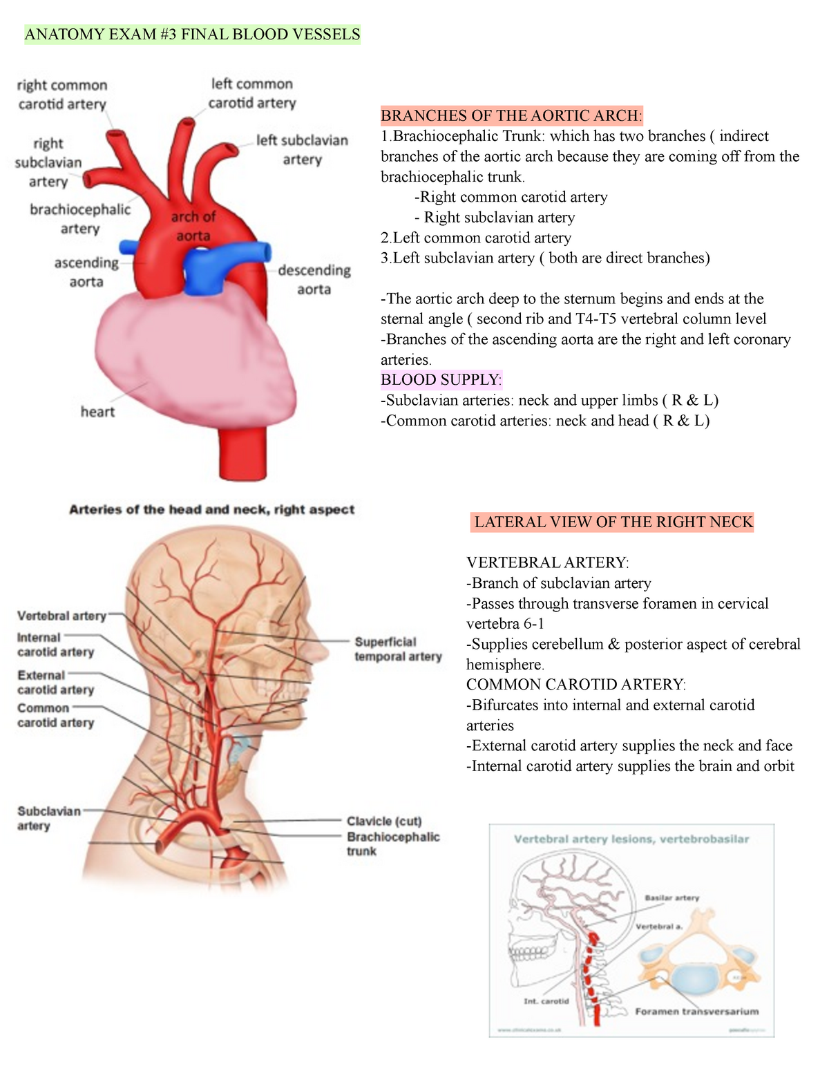
Anatomy Test 3 Anatomy Exam 3 Final Blood Vessels Branches Of The Aortic Arch 1 Trunk Which Has Studocu
Comments
Post a Comment